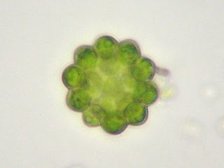
This is an image that I posted last week. I returned to this alga to find out more about it. I read through some books and identified it as a Coelastrum microsporum (Bold & Wynne). It is a multicellular, green algae in the Chlorophyta division.

This is the Phacus sp. I photographed and video recorded in the past couple of weeks. I included these photos because they show different angles of its body.


The organism below is a Euglena sp. part of the phylum Euglenophyta (Forest). It has a red "eye spot" that is sensitive to light. It is located near the base of its flagella, seen in the photo towards the left end of the organism.

 This is another example of a flagellate organism. I identified it as an Entosiphon. It is also in the phylum Euglenophyta (Patterson). It has two flagella, one on either end. Only one is visible in the photo to the right, as the second is very small.
 The photo to the left shows two cyanobacteria (Forest). The large bulb-like end on each is a heterocyst. Heterocysts are thick-walled parts of cyanobacteria that have the ability to fix nitrogen.
The following video and photos show examples of larger organisms in my MicroAquarium. The video below shows a Philodina sp. It is a kind of Rotifer (Pennak).
 To the right is an example of an Ostracoda. This organism has a more common name of Seed Shrimp (Smith). This is the largest organism I have found in my aquarium thus far.
 This is an excellent photo I was able to capture of a premature Cyclops. These organisms move very quickly and are difficult to photograph.
The organism below is of the genus Halteria (Pennak). It also move very quickly, and is hard to photograph. They are certainly smaller than the Cyclops and the Seed Shrimp. They are closer in size to a shelled rotifer.

 I ended my blog last week by discussing a newly observed organism. This week I saw several more examples of this organism. Many of them were near the base, and in the sediment. However, I still observed some swimming through the open water of my aquarium this week. The organism to the right is called a Gastrotrich (Patterson).
Bold H.C. & Wynne M.J. Introduction to the Algae, Structure and Reproduction. 2nd ed. Englewood Cliffs (NJ): Prentice-Hall, Inc. p. 148.
Forest H.S. 1954. Handbook of Alga. Knoxville (TN): University of Tennessee Press. p. 274-427.
Patterson D.J. 1996. Free Living Freswater Protozoa, A Coulour Guide. Lonon (UK): Manson Publishing Ltd. p. 28-53.
Pennak R.W. 1989. Freshwater Invertebrates of the United States 3rd ed. New York (NY): John Wiley & Sons, Inc. p. 81-172.


